
For almost every organ in humans there is a match in flies, and common genes regulate their development, organisation and function.
Introduction
Living animals have fundamental physiological requirements. Just like us, flies have to breath, digest, move, learn and coordinate in order to survive. Due to our shared evolutionary history, many of our organs have common origins and serve the same purposes, and their development, organisation and function are often regulated by the same genes.
Deciphering the principal functions of these genes and the developmental and physiological processes they regulate, can be done efficiently and cost-effectively in Drosophila, as is explained under the “Why fly?” tab. Applying this new knowledge from Drosophila to human organs in health and disease is a powerful, much used and well proven strategy which inspires and accelerates research in higher animals and humans – often referred to as using the fly as a “test tube”. On this page, the most important organs and tissues are briefly described and compared – a helpful resource for schools and also for universities. Another excellent resource is the book by L. I. H. Held Jr. “Deep homology? Uncanny similarities of humans and flies uncovered by evo-devo” (2017, Cambridge University Press, Cambridge) — [LINK1] [LINK2].
If you want to learn about how these organs are formed during the development of the fly, please visit Volker Hartenstein’s Atlas of Drosophila development, or the FlyMove/Organogenesis page.
Body organisation
As illustrated by different colours in the image below, the bodies of humans and flies are subdivided into segments. When looking at our bodies from the outside, this segmentation might not be obvious at first sight. However, it becomes clear when seeing the segmental nerves which emanate from our spinal cords in a regular pattern (one pair per segment; see image of the next section) or considering the vertebrae in the human backbone which each reflect one segment. These segments are organised into larger structural units, and in humans and flies we can distinguish a head, thorax and abdomen. Although the appendages of humans and flies differ (2 arms and 2 legs versus 6 legs, 2 wings and 2 halteres) they share the common principle of being located in stereotypic positions of only a small number of specific body segments.

Flies and humans show a segmental anatomy (different colours) subdivided into head, thorax and abdomen
The subdivision of our bodies into segments, their organisation into head, thorax and abdomen, as well as the precise location of appendages are regulated through precise developmental processes referred to as patterning mechanisms. See in the following film how these processes work in the fly embryo:
——————— back to top ———————-
Nervous system
As the example of segmental nerves illustrates, also the nervous system is subdivided into segments. In flies and humans, segmental nerves contain:
- motor nerves conduct information from the central nervous system (CNS) to muscles and glands. These nerves are often affected in motorneuron disease leading to gradual paralysis (see explanatory images here).
- sensory nerves conduct information from sensory organs to the CNS. These nerves (together with motor nerves) are affected in Charcot–Marie–Tooth disease leading to gradual muscle wastage due to lack of usage.
In both organisms, cell bodies of the nerve cells (neurons) constituting sensory nerves, lie outside the CNS: in humans primarily in dorsal root ganglia (red in image below), in flies within the sensory organs at the body surface (see illustration). In both organisms, sensory organs are for vision, smell, taste, hearing, balance, mechanical information, temperature, noxious stimuli etc.
Flies and humans show a subdivision of the CNS into brain (in the head region) and ventral/spinal cord (in the trunk). Also the smallest functional units, the nerve cells, have much in common in humans and flies, and the same genes equip them with synapses and the ability to fire action potentials. Many fundamental mechanisms about their development and function have been discovered in flies, including mechanisms of neurodegeneration (more info here). Our behaviours are coordinated through the nervous system and, even at this level, we share certain commonalities (see examples of alcohol-related behaviours here).

Organisation of the nervous system into brain in the head, ventral/spinal cord in the trunk and segmental nerves
In flies and humans, the ventral/spinal cord primarily contains neuronal networks which regulate local events within the trunk, often within a single segment, and a good example are motor reflexes. In contrast, the brain connects to all segments and acts as a coordinating centre for the whole body. Accordingly, the brains of flies and humans are highly specialised and subdivided into functional centres for vision, smell, motor coordination, learning and many other behaviours. A more detailed descriptions about the sensory centres is given in our L3-Neurons resource.

The brain is organised into functional centres as colour-coded for human and fly brain.
To see more illustrations of the Drosophila nervous system, just browse here. To see a comparison of human and fly eyes, see our L5-Vision resource.
IMPACT: Work on the nervous system of Drosophila has made seminal contributions to modern neurobiology (Bellen et al., 2010, Nat Rev Neurosci 11, 514ff.). To name but a few: (1) It has delivered the first genes underpinning action potentials (still studied in epilepsy research). (2) It has given us understanding of synaptic genes and fundamental mechanisms of neuronal stem cell regulation. (3) It pioneers mechanisms of nervous system wiring during development. (4) It made major contributions to the understanding of how nerve networks form the cellular basis of learning and behaviour (see Virtual Fly Brain) which delivers highly informative data, strategies and techniques also for the Human brain project. (5) It provided the first learning genes, (6) the molecular mechanisms of our biological clock, as well as (7) understanding of gene functions in neurodegeneration (see here), (8) the role and regulation of sleep, (9) the physiology of vision, and (10) it led to a Nobel Prize for important discoveries on the cellular and genetic basis of the smell sense.
——————— back to top ———————-
Moving
Movement in/of our bodies depends on the contractile forces of our muscles:
- cardiac muscles are the muscles that line the heart wall and are different from other muscles because they have neuron-like electrical properties, both in humans and flies (see circulation);
- smooth muscles move soft tissues in our bodies, such as the peristaltic movements of our gut, or the constriction of blood vessels which regulates blood pressure; also the gut of flies is lined by smooth muscles;
- skeletal muscles are the only muscles that are under voluntary control and serve to move our bodies and enable us to breath; they anchor with their tendon tips to bones which are therefore moved upon muscle contraction; in contrast to smooth muscles, skeletal muscles appear striated under the microscope in both flies and humans, because the contractile filaments in their cores are organised into periodic, regularly sized and spaced blocks that occur like alternating stripes on a football scarf (see images below). In both organisms, skeletal muscles are the product of many precursor cells fusing into one large muscle fibre that contains many cell nuclei. In the small fly one muscle fibre is the equivalent of a muscle, whereas the large muscles in humans are composed of many muscle fibres. However, the way in which muscles work are identical (see film below or an alternative video here).

Skeletal muscles of humans and flies are structurally very similar.
As should have become clear, the muscles of humans and flies share a lot in common at the molecular and structural level. In contrast to muscles, the skeletons of humans and flies are very different from one another:
- humans have an endoskeleton in form of bones which are embedded deep in our bodies and consist primarily of crystallised sugary proteins, called type I collagens;
- flies have an exoskeleton deposited on the body surface in form of cuticle (see image below); the key components of cuticle are specific fibrous sugars called chitins, but there are further components including wax and various sugary proteins.

Although the location as well as the constituents of bones and cuticle are very different, they have in common that they represent extracellular matrix (ECM; latin “extra” = outside). ECM is composed of mostly structural molecules deposited outside cells. Therefore, bones and cuticle are are primarily dead materials which, consequently, persist as skeletons when our bodies decompose. ECM is made, released and modulated by specialised cells. In the case of human bone, ECM is produced by osteoblasts embedded in the matrix, and cuticle is made by skin cells (see red arrows in figure above).

Although this fly skeleton looks rather cool, it is unfortunately only science fiction!
Skeletal muscles do not attach directly to the skeleton, but they are linked through specialised ECM structures, called tendons in humans and tendon matrix in flies:
- tendon is composed of fibrous type II collagens and deposited into need parallel bundles by dynamic cells called tendon fibroblasts (see image above); at one end, tendons link to muscle surfaces (myotendinous junctions), on the other to the ECM of the bone (enthesis).
- tendon matrix in flies is composed of different types of extracellular proteins which are deposited by the skin, muscle cells, and motile cells in the body fluid called haemocytes. Like in humans, the fly tendon matrix links to muscles on one side. On the other side, it does not link directly to the skeleton but to the surfaces of skin cells. These skin cells form a dense intracellular (latin “intra” = within) array of skeletal elements linking the tendon junction to the cuticle junction on the other side where they anchor via specific extracellular filaments called tonofilaments (see image above).
The cuticle in juvenile fly maggots is softer than in the adult fly, and muscle contractions lead to a movement that is comparable to peristaltic waves in the gut – as you will have similarly observed in earthworms or caterpillars. It is shown for a Drosophila maggot in this fist movie below. The second movie shows the corresponding activity running in a wave along the “spinal cord”, called ventral nerve cord, of the maggot. For a schematic presentation of ventral nerve cord and muscles see this image.
——————— back to top ———————-
Breathing
To generate energy, the cells in our bodies perform complex reactions in their mitochondria which use oxygen (O2) and generate carbon dioxide (CO2) as a waste product (explained here). O2 supply and CO2 disposal in humans is provided through the lung, in many aquatic animals by gills, in most arthropods including flies through the tracheal system.

Lungs and tracheae are highly branched, tubular structures which are filled with air.
The lung is a highly branched, tubular structure which is directly connected via its main branch (the trachea) to the mouth opening. Within the lung, the hollow air-filled trachea branch out into fine bronchioles which terminate in balloon-shaped alveoli (singular: alveolus) which are grouped or bunched up into acini (singular: acinus; see film below). Alveoli are closely associated with fine capillaries of the blood circulation system. This close spatial arrangement allows O2 to pass over from the hollow alveoli into the blood, which is carried via pulmonary veins (red in image) to the left heart chambers (see “Circulation” section) and from there to all parts of our body, where the O2 can be passed on to target cells in need. Vice versa, the pulmonary arteries (blue in image) carry the CO2-rich blood from the right heart chamber into the lung, where the CO2 is released into the alveoli to be exhaled during the normal breathing process.
The tracheal system of flies also consists of tubular structures connecting to the body surface at one end, and branching out into the body on the other. Flies are small and do not have a blood vessel system that enters tissues. Instead, the tubular tracheal branches reach directly into all organs and tissues and exchange O2 and CO2 directly with their cells (see image above).
The process through which O2 and CO2 pass over between the different cells (lung, tracheae, blood, target cells) is called diffusion. Diffusion is the passive flow of molecules from places of high concentration to low concentration. For this, the air we inhale contains ~21% of O2 (a good source relative to arterial blood or fly tissues) and 0,04% CO2 (a good sink relative to arterial blood or fly tissues). In addition, human blood contains red blood cells (erythrocytes) which contain the red hemoglobin bio-molecules that very efficiently bind O2. This raises the capacity of blood to transport O2 by 70-fold. When erythrocytes are carried into areas where cells are in need of O2, the CO2 levels are usually high, and this leads to an increase in acidity (as you might know from drinking fizzy water!). The increase in acidity triggers the release of O2 from hemoglobin, which is now free to diffuse into the target cells.
See here a cool movie showing how the tracheal system develops in a Drosophila embryo:
——————— back to top ———————-
Circulation and blood
Humans and flies use circulating body fluids to deliver necessary substances such as nutrients as well as hormonal messages to cells, to transport metabolic waste products away from cells, and to provide a medium through which blood cells can move around and police the body. In humans also oxygen and carbon dioxide are transported via the blood and red blood cells, whereas insects have developed a tracheal system (see Breathing). The circulating body fluids of humans are called blood and lymph, whereas there is only one fluid called haemolymph in flies:
- The blood is pumped by the heart and circulates through the closed blood vessel system: it flows away from the ventricles of the heart through thick arteries which branch into thinner arterioles and further into the networks of very fine capillaries in the target tissues (either lung or other body tissues), from where it flows off through thin venules which merge into the thicker veins that eventually lead back into the heart’s atriums.
- The lymph fluid is recollected liquid from tissues of our bodies, primarily generated through spill from blood vessels. Lymph flows off through vessels of the lymphatic system which acts as a drainage system that eventually leads into the veins close to the heart, thus re-uniting lymph and blood.
- Flies have only one circulating body fluid which is consequently termed haemolymph. It fills the whole body of insects encapsulated only by the epidermis (equivalent to our skin), and all organs and tissues inside insects are emerged in this fluid. This liquid is circulated via a tubular heart which acts like an aquarium pump: it sucks in water from the posterior body end and releases it in the anterior body (or vice versa: see below), thus gradually circulating the whole body volume of haemolymph (see image).

Let’s briefly compare the heart anatomies. The human heart (see image) is rather complex in that it is subdivided into two halves:
- the right side sucks in blood from the head and body to then pass it on to the lungs (see “Breathing” section),
- the left side receives blood from the lungs and pumps it into the head and body.
This rather complex organisation has evolved from more primitive heart versions as they are still present in fish. The fish heart is organised into a sequence of bulgy chambers which are folded into a Z-like shape (see image), but it is still recognisable as one continuous canal. This structure is far easier to compare to the heart of Drosophila which is divided into three parts.
- the back part consists of a tube that performs peristaltic contractions (see movie below) to suck in/release haemolymph from/into the abdomen of the fly;
- the central part is the conical chamber which is bulgy and reminds of a ventricle;
- the anterior part is the aorta which extends the most important haemolymph exit point to the distant end of the body, thus ensuring circulation along the entire length of the fly (see image).
- The fly heart primarily pumps body fluid from the abdomen to the thorax, but it has been reported (see here) that this direction of flow can be reversed. For this to happen, the fly heart contains two different sets of valves:
- Ostias are 5 pairs of openings towards the haemolymph (1-5 in image) which allow body fluid to enter but not exit the heart tube. When haemolymph is pumped from back to front, all ostias jointly allow entry from the middle and hind part of the body, which is then pumped through the aorta towards the front (light blue arrows). When in reverse mode, the ostia pair 1 in the conical chamber allows entry of body fluid from the middle of the fly which is then pumped out via the aorta at the front as well as through the heart tube opening at the back end of the heart (light orange arrows).
- Valves are specialised cardiomyocytes which can close off the heart tube at different locations, and they perform this flow-stopping actions well synchronised with the peristaltic heart contractions, thus ensuring a clear direction of flow either to the front or the back opening, depending on the mode.
As indicated in the picture, there are alary mucles associated with the heart which are likely to give the heart lateral stability and support. The following movie shows a heart beating in a larva, and the YouTube video below in an adult fruit fly:

Obviously the human and fly heart serve similar purposes, and the heart cells (cardiomyocytes) which form the walls of the human and fly heart share a number of evolutionary well conserved physiological properties:
- As indicated in the muscle section, heart muscle cells of both flies and humans have electrical properties reminiscent of nerve cells, and this allows them to beat autonomously (i.e. without being excited by nerve cells). To regulate the heart beat, both flies and humans have pacemaker cells at either end of the heart.
- Furthermore, heart muscle cells of flies and humans use very similar sets of genes for their development and function. For example, flies and humans share a highly specific ion channel proteins required for heart beat regulation that the faster beating mouse heart does not have.
Also blood cells have commonalities between humans and flies, and their development as well as function partially depends on very similar genes:

- In humans, new blood cells are constantly produced in the bone marrow, split into two major lineages:
- The lymphoid lineage gives rise to lymphocytes, i.e. the B- and T-cells constituting the adaptive/acquired immune response. They form an armada of individual cells, each randomly specialised via a key-in-lock mechanism to recognise specific cues on invading pathogens. If such a pathogen enters our body, the appropriate lymphocytes get activated, meaning they will multiply and either attack and destroy the invader or generate specific antibodies that will bind and tag the pathogen. The adaptive response system involves a number of organs: the thymus (where accidental lymphocytes are eliminated that would otherwise attack our own bodies), the lymph nodes (where dendritic cells present detected pathogens collected in the body periphery, in order to identify and activate those lymphocytes which can fight the invader), and the spleen (where antibodies get produced large scale and the blood is filtered for pathogens which are bound by antibodies). The adaptive immune response is specific to vertebrates and cannot be found in flies.
- The myeloid lineage produces red blood cells (erythrocytes) for oxygen transport (not found in flies), as well as various cell types which represent the more ancient innate immune response, and these cells have clear similarities to blood cells of flies (see image above). These cells are key to any immune response, since they represent the first and fast defense system: (1) they can attack straight away (through chewing invaders up, releasing anti-microbial substances, or weaving molecular nets called extracellular traps in the infected area to catch them), (2) they produce signalling substances (cytokines, that draw in more immune cells to join the defense), (3) they pass this information on to lymphocytes to activate the second defense system, i.e. the adaptive response (not present in flies).
- In flies new blood cells are generated in haematopoietic hubs associated with the heart (see first image above). Fly blood cells comprise plasmatocytes or macrophages (engulfing cell debris, bacteria and fungi), crystal cells (involved in melanisation response and wound healing) and lamellocytes (large cells which can engulf large invaders, such as eggs of parasitic wasps). Plasmatocytes and crystal cells are derived from progenitor cells in the haematopoietic hubs, whereas lamellocytes seem to be derived from plasmatocytes.
In order to fulfill their functions, blood cells need to move around our bodies. See in this fascinating movie how blood cells (haemocytes) move through the Drosophila embryo:
——————— back to top ———————-
Digestion
The alimentary canal of humans and flies has a similar organisation into foregut, stomach, mid- and hindgut. Also some of the accessory glands associated with the human alimentary tract have (partial) matches in the fly:
- the salivary gland produces saliva for pre-digestion and has an obvious match in the fly;
- the liver with its various functions (including detoxification and the production of enzymes necessary for digestion) has its closest match in the fat body which is spread out widely in the fly abdomen (grey in image);
- the pancreas produces digestive enzymes but is also a hormonal gland; clusters of cells called islets of Langerhans produce insulin and find their match in a group of cells located in the fly brain (yellow in image).

Colours indicate homologous and functionally related structures of the alimentary canal.
As is explained in the next section, the border region between hind and midgut harbours the exit openings of the Malpighian tubules releasing their urine into the gut, whereas in humans the gut and ureter are clearly separated.
——————— back to top ———————-
Excretion
Humans and flies produce urine as a means of detoxification which, in humans, is performed by kidneys and in flies by Malpighian tubules (also depicted in the digestion section). Both organs display excretory tubules designed to selectively filter toxic substances from the body fluid (blood in humans, haemolymph in the fly) which are disposed of via the urine, whilst retaining the “good stuff” and as much water as possible within our bodies. As shown recently [LINK], Malpighian tubules in Drosophila even form kidney stones as a function of genetic predisposition.
In humans, the filtration of toxic or waste substances from the blood is performed in the glomerulus (also called Bowman’s capsule) by specialised cells called podocytes. The homologous cells in the fly are called nephrocytes which are found in the thorax as Garland cells and the abdomen as pericardial cells. Nephrocytes have a structurally very similar filter apparatus which has a well conserved genetic basis. Defects in some of these related genes lead to a disease called proteinuria (high proteins levels in the urine) in both humans and flies. In contrast to podocytes which can excrete waste products via the urine, nephrocytes in Drosophila have no access to direct excretion and are often referred to as storage kidney.
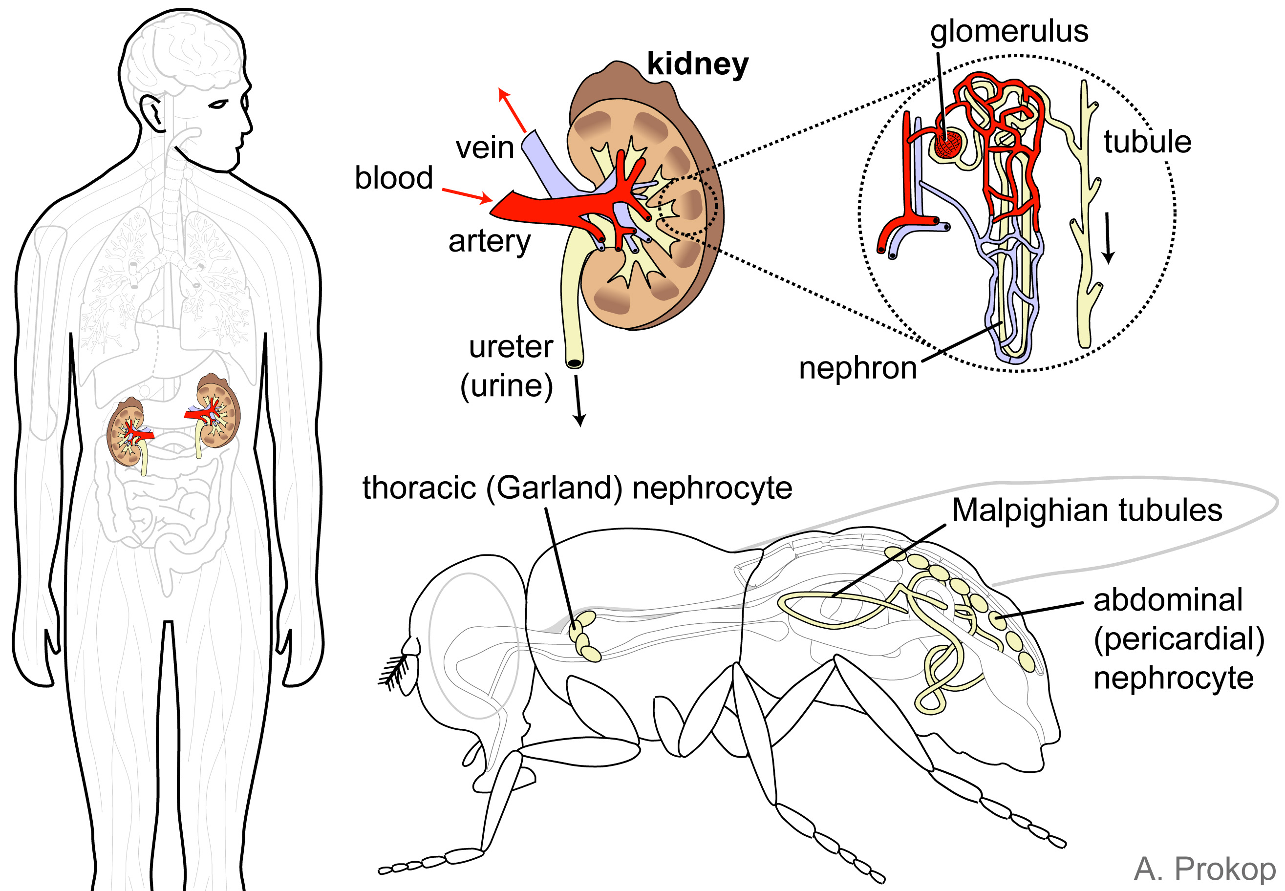
Kidney in humans versus nephrocytes and Malphigian tubules in flies, filter toxic substances from our body fluids
——————— back to top ———————-

Thank you so much for this. Great article; I’ve already sent it to several colleagues and labs.
LikeLike
Thanks a lot, Rami. You might also like our other site (http://www.flyfacility.ls.manchester.ac.uk/forthepublic/), as well as two recent blogs: https://poppi62.wordpress.com/2015/05/12/fly-scicomm/ and http://blogs.brandeis.edu/flyonthewall/why-do-we-have-to-learn-this-stuff-establishing-drosophila-as-a-modern-teaching-tool-in-schools/
LikeLike
These images are great! I was hoping to use them in a talk I am giving soon to a bunch of ‘non-fly’ people. Can I reference them to this website or is there a more appropriate reference for them?
LikeLike
Pingback: Bringing life into biology lessons: using the fruit fly Drosophila as a powerful modern teaching tool | Gedankenexperimente
Pingback: Bringing life into biology lessons: using the fruit fly Drosophila as a powerful modern teaching tool | Pedagoo.org
Pingback: How flies are making their way into classrooms | Faculty of Life Sciences, UoM
great images for teaching
LikeLike
Andreas: a fantastic overview of organ system comparison and similarities. I was looking of a nice image depicting human and fly for a talk and found your images really perfect.
Regards
Simon
LikeLike
Thanks for the kind words, Simon. Please, spread the news also about the other resources on this website as well as on our other website: http://www.flyfacility.manchester.ac.uk/
LikeLike
Hi Andreas,
Your material is fantastic, we have used it many times during school visits to our institute.
I would like to point to a minor mistake in one of your images. The spleen has been placed on the right side of the body, while it is actually on the left.
thanks for all the hard work that helps to spread the benefits of flies as a model organism
LikeLike
Thanks for the heads-up. Hope to find some time to correct that.
LikeLike
How do we cite the diagrams ?
LikeLike
Manchester Fly Facility (2015). droso4schools: Online resources for school lessons using the fuit fly Drosophila — https://droso4schools.wordpress.com/
Thanks for asking!
LikeLike
Great article. Very well put together and this is really helpful and reliable. Thanks for sharing this and keep up the good work. Very much appreciated.
LikeLike
Pingback: Science communication in the biomedical sciences: challenges, opportunities and new approaches | Gedankenexperimente